loader

Request Appointment

Error: Contact form not found.

Month: August 2026

7August 2026

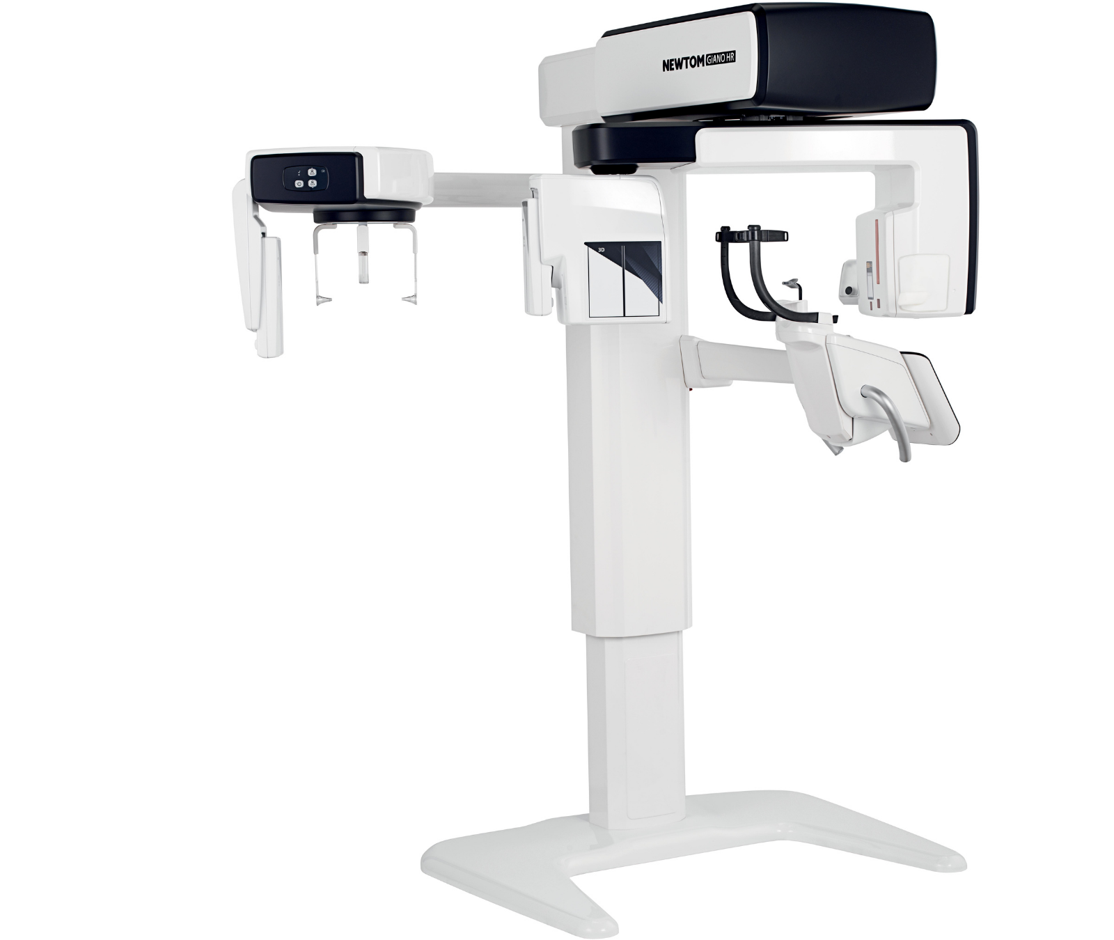
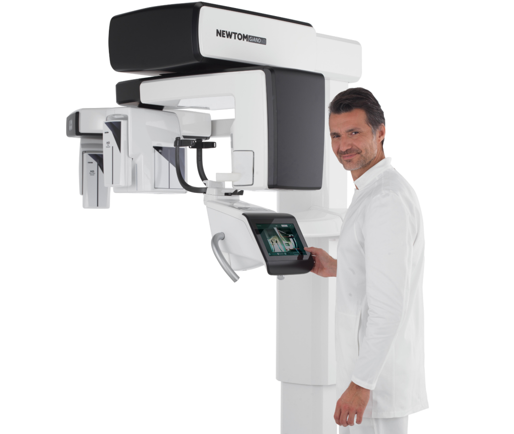
GiANO HR FullView

Complete radiology solution.


Five FOV configurations for different diagnostic fields

  • Essential Configuration

    NEWTOM GiANO HR FullView comes, as standard, with a range of FOVs wide cover multiple treatment areas: endodontics, implantology, orthodontics and general dentistry.


    FOV: 6×6 – 8×6 – 8×8 – 10×6 – 10×10 – 11×8 – 13×6 – 13×10

  • Superior Configuration

    Together with the Essential configuration, the Superior FOV range extends the scope for treatment to the gnathology, otolaryngology and the maxillofacial field, including scans of both adult temporomandibular joints.


    FOV: 13×16 – 15×6 – 15×10 – 15×16
    FOV: 17×10 – 17×14 – 17×18

  • TMJ Pack

    For high-definition analysis of the inner ear and petrous bone, and any dysplastic, inflammatory or trauma-related pathologies affecting the cervical spine.


    FOV: 15×6 – 15×10

  • Cervical & Ear Pack

    For high-definition analysis of the structures of the inner ear and petrous rock, and any dysplastic, inflammatory, and traumatic pathologies that may affect the cervical spine.


    FOV: 7×6 – 9×9 – 9×16

  • ENDO Pack

    For endodontics and implantology analysis at the highest resolution, reaching 50 μm (Voxel).


    FOV: 4×4 – 5×4

2D Ultra HD Diagnostics

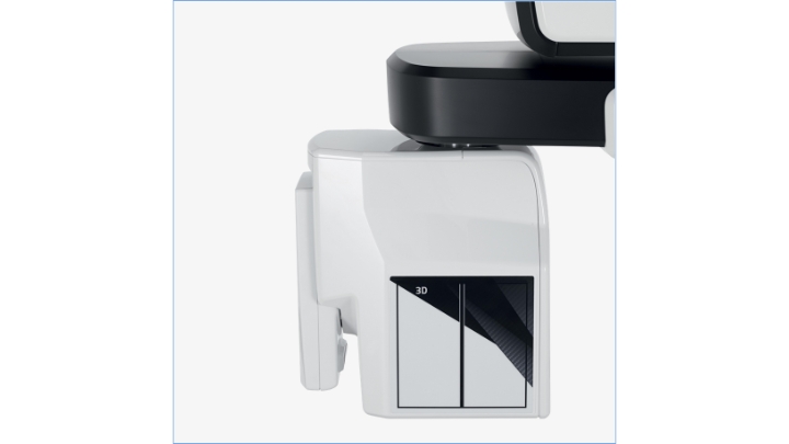
Unlike traditional sensors, the Direct Conversion Detector does not require conversion of X-rays into visible light as it is capable of sensing it directly and converting it into precise, accurate digital signals.

In this way, extremely high resolution diagnostic images can be obtained at low X-ray doses, and highly detailed images can be produced even where quick scans in ECO Dose are more suitable.

Advanced software

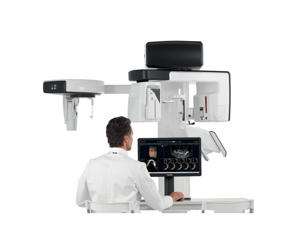
Neowise integrates automated AI-powered features that improve diagnoses, raise operational efficiency and make treatment more personalised for each patient, making your work more precise and finely targeted than ever

  • Detection of panoramic curves on CBCT scans
  • Identification of inferior alveolar nerve in volumetric scans
  • 2D and 3D Data Classification
  • Anatomical and pathological analysis for 2D intraoral and panoramic scans
  • Segmentation of 3D anatomical structures
  • Airway identification in cephalometry for the diagnosis of OSAS pathologies
  • Alignment of latero-lateral teleradiography with photo of patient
  • Alignment and combination of CBCT scans with optical impressions
  • Detection of cephalometric points and creation of tracings
  • Smile Design module to simulate aesthetic treatments in frontal sectors